The Anatomy Of The Neck: Bones And Cartilage
The anatomy of the neck is very demanding, as numerous structures are housed here in a very small space.
When we look at the anatomy of the neck, we can understand a little bit more about our body. The neck is the part of the body that acts as a bridge between the torso and the head. It is a vital structure through which important blood vessels and nerves run.
It’s a thin and flexible area that allows us to move our head. However, it is also a sensitive area as any injury can damage the blood supply to the brain or its nerve connections.
The neck is more complex than it seems at first glance. It consists of a number of bones, cartilage, muscles, vessels and nerves. So, in this article, we’re going to focus on the bones and cartilages that make it up!
Neck Anatomy: How Can We Define It?
To make examining the neck a little easier, specialists have put a number of superficial boundaries to define it. The neck begins at the bottom of the jaw and the occiput, which forms the base of the skull. At the front it then extends to the collarbones and the sternum. In the back, the neck reaches the C7 vertebra.
The neck is one of the most complex structures in the human body because it contains many important elements that come together in a very small space, such as:
- The carotid artery
- The pharynx
- The larynx
- The trachea
- Numerous nerves
Now let’s talk a little about the outer framework that protects all of these body parts.

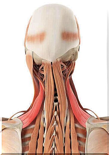
The anatomy of the neck: what bones do we have in our neck?
When we talk about the anatomy of the neck, we cannot ignore the bones! The skeleton of the neck consists of the cervical vertebrae, the hyoid bone, the collarbones and the sternum. Seven vertebrae form the cervical part of the spine, i.e. the neck.
In addition, there are intervertebral joints that give it flexibility and movement. In fact, this structure is sensitive to shocks and occasionally causes pain.
Cervical spine
The vertebrae are different. As we mentioned earlier, the neck is made up of seven vertebrae. From the third to the sixth they are all the same:
- They have a vertebral body and a spinous process – this is the back part of the vertebra.
- They have a concave top and a convex bottom.
- They are also small and a little flattened in relation to the rest.
The first vortex is called the atlas. It is a kidney-shaped bone, without a body or apophysis. This vortex is formed by two lateral masses connected by arcs, hence its ring shape. It is the vertebra that touches the occiput.
The second vertebra, or C2, is the axis. What sets it apart from the rest of the cervical vertebrae is its process. It’s called the odontoid process and is a protrusion on your body in the shape of a pencil.
The C7 vertebra also differs from the other vertebrae by its extension. It is a spinous process, like the C3 to C6 vertebrae, but it is not bifurcated. This spinous process is longer than the other cervical vertebrae and is also referred to as protruding because of its special shape.
Hyoid bone
This is a movable bone that is located in the front part of the neck. It lies at the level of the third vertebra, between the jaw and the thyroid cartilage. In addition, the hyoid bone is not connected to any other bone. It is held in place by a series of ligaments that lead to the temporal bones of the skull, called the styloid ligaments. In addition, it is anchored to the thyroid cartilage.

Cartilage and ligaments of the neck
First of all we have to mention the thyroid cartilage, the cricoid and the epiglottis. They form the front part of the neck, they are also part of the larynx and enable us to breathe.
On the other hand, the intervertebral discs between the vertebrae form the joints of the cervical spine, which are connected by a series of ligaments. The intervertebral discs have a central part, the nucleus pulposus, and an outer part, the annulus fibrosus.
The anterior longitudinal ligament connects the vertebrae in the anterior region. There is also a rear longitudinal band. In addition, the yellow band connects the joints of two vertebrae, followed by their back.
The intertransverse ligament and the interspinous ligament connect the vertebrae between their apophyses. Finally we find the ligamentum supraspinatus, which at its highest point forms the ligamentum nuchale.
Conclusion
Now that we’ve learned about the anatomy of the neck, I think we can all agree that it is a very complex part of our body. To understand how our throat works, we need to study it in depth. The seven vertebrae with the associated ligaments and intervertebral discs ensure that the neck is flexible and the head can move.