Interesting Facts About Multiple Pilomatrixomas
Multiple pilomatrixomas usually occur in myotonic dystrophy or an acquired genetic mutation. But other diseases can also lead to it.
Multiple pilomatrixomas are very rare. It is a benign tumor in the hair matrix that normally occurs sporadically. The multiple form is extremely unusual.
There are no precise data on the incidence of this type of tumor. Because not much is known about the pilomatrixoma in its typical form. Often this disease is confused with other pathologies.
What is known is that around 2 to 3.5% of total cases are multiple pilomatrixomas . Usually there is a family history. Learn more about this rare disease today.
Multiple pilomatrixomas: what is it?
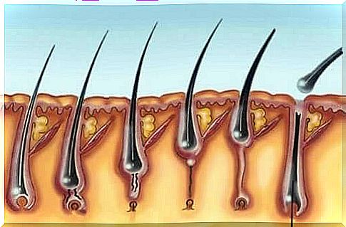
A pilomatrixoma is a benign tumor in the hair matrix that develops for reasons that are as yet unknown. It usually happens in childhood. It is estimated that 10% of skin cancers in children are of this type.
This tumor develops in the subcutaneous tissue and often extends to the fatty layer of the skin. If several such tumors are present, one speaks of multiple pilomatrixomas. These are mostly due to hereditary factors or other diseases.
40 cases are currently associated with a genetic predisposition and 30 cases with a muscle disease called myotonic dystrophy or Curschmann-Steinert’s disease. Some scientists believe it is just a symptom of myotonic dystrophy.
However, multiple pilomatrixomas could also be found in connection with other diseases. For example, they can occur in Gardner syndrome, Rubinstein-Taybi syndrome, medullary thyroid carcinoma (MTC), a trisomy, mild coagulation disorders or complications after a sternotomy. There are also patients in whom there are no associated abnormalities.
Multiple pilomatrixomas: characteristics
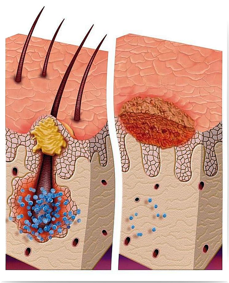
A pilomatrixoma can be recognized by a hard lump that is usually the same color as the skin. This can happen at any age. But 60% of the cases occur in the first 10 years of life. There are also more cases between the ages of 60 and 70, with women slightly more likely to be affected.
Over 50% of those affected have the lumps in the head and neck area. But they can also appear on other parts of the body with hair follicles. If the lumps are superficial, they turn the skin bluish-red in color. A hard core can be felt when scanning.
As already mentioned, multiple pilomatrixomas can occur in myotonic dystrophy. It is a muscular dystrophy that is genetically determined and in which there is a progressive breakdown of the muscles (muscle weakness). In each subsequent generation, this disease is more intense.
Multiple pilomatrixomas can lead to different injuries, sometimes occurring at the same time, forming a type of plaque. The nodes develop slowly and asymptomatically. Usually the patient himself discovers the lump by palpation.
Multiple pilomatrixomas: causes and diagnosis
Multiple pilomatrixomas are believed to be related to a group of proteins called catenins. These are responsible for connecting the actin component of the cytoskeleton with cadherins. Everything indicates that a mutation in beta-cetenin is the underlying problem. This could be confirmed in 75% of the pilomatrixomas.
The clinical symptoms of this disease are those of myotonia (muscle disorders). Muscle weakness and atrophy develop. In addition, opacities of the lens, slight cognitive impairments, premature balding in the front area, testicular atrophy, heart block and abnormalities in electromyography occur.
The diagnosis is made through clinical examinations. In some cases, an ultrasound scan is necessary. Medical history, a hematogram, and urine and stool analyzes are also necessary to confirm the disease and identify or rule out possible associated syndromes.
useful information
Sometimes the nodes are very fine, atrophic and uneven or resemble a bubble. However, this is only the case in 2% of cases, especially in young women in the area of the extremities.
In very rare cases, a crust similar to an ulcer develops . However, only 11 cases of this type are known. One then speaks of a perforating tumor.
Simple and multiple pilomatrixomas are identical. But there are four cases in which multiple pilomatrixomas are associated with macular degeneration or anetoderma. After a pilomatrixoma is diagnosed, it must be observed over the long term. It is usually removed by surgery.